Upper Leg Tendon Anatomy / Anatomy Of Leg Muscles And Tendons Lower Leg Anatomy ... / Hands are outstretched, holding onto the handles of the bench.
Upper Leg Tendon Anatomy / Anatomy Of Leg Muscles And Tendons Lower Leg Anatomy ... / Hands are outstretched, holding onto the handles of the bench.. Tendon, tissue that attaches a muscle to other body parts, usually bones. Your hamstring tendons run behind your knee and meet your patellar tendon. Upper legs anatomy — stock image. See the pictures and anatomy description of knee joint bones, cartilage, ligaments, muscle and tendons with resources for knee problems & injuries. Medically reviewed by william morrison, m.d.
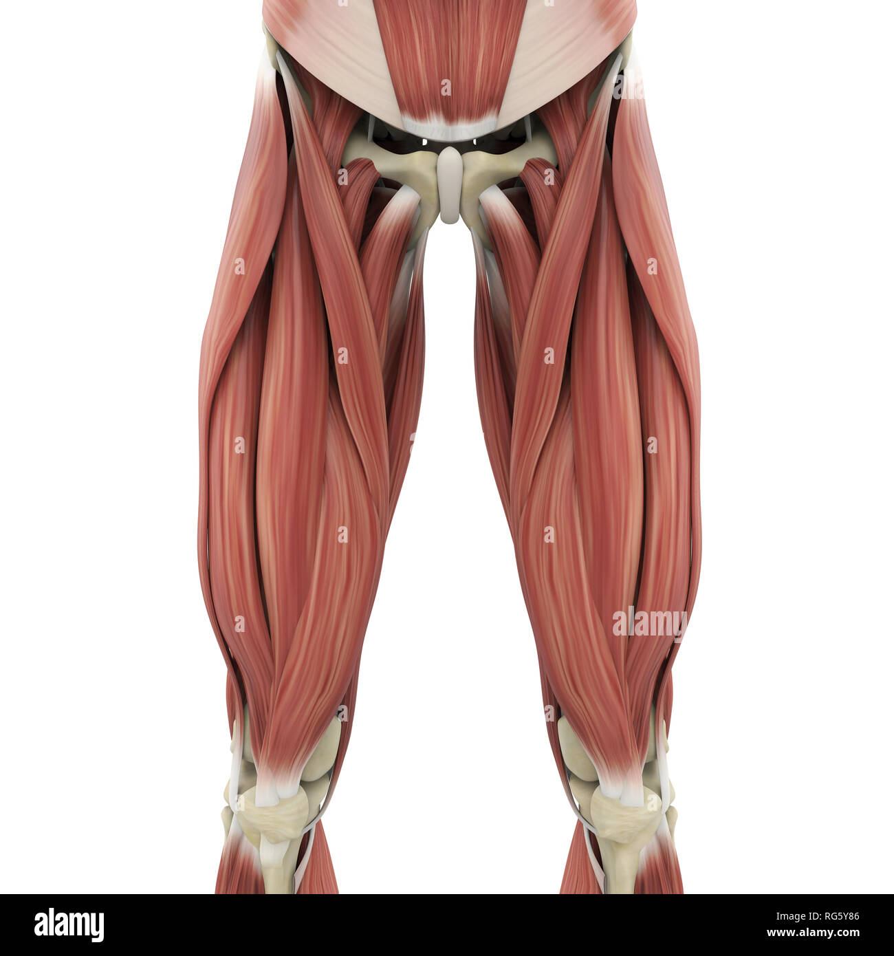
Tusindvis af nye billeder af høj kvalitet tilføjes hver dag. It's the area that runs from the hip to the knee in each leg. There is no real division between the core and the upper leg; They are remarkably strong, having one of the highest tensile strengths found among soft tissues. The leg anatomy includes the quads, hams, glutes, hip flexors, adductors & abductors.
Human Upper Leg Muscles High Resolution Stock Photography ... from c8.alamy.com Peroneal tendonitis affects these tendons, and can make movement difficult and painful. Superficial veins of upper limb , anatomy : Study upper leg anatomy flashcards from tony hao's university of leicester class online, or in brainscape's iphone or android app. Medically reviewed by william morrison, m.d. Upper legs anatomy — stock image. The peroneal tendons are in the feet and provide balance and stability during movement. Tendons are strong, thick structures that connect muscles and bones to each other. Collectively, they act to dorsiflex and invert the foot at the ankle joint.
Find stockbilleder af concept 3d human upper leg anatomy i hd og millionvis af andre royaltyfri stockbilleder, illustrationer og vektorer i shutterstocks samling.
Related online courses on physioplus. The achilles tendon (tendo calcaneus or tendo achillis) is the thickest and strongest tendon in the human body. The human leg, in the general word sense, is the entire lower limb of the human body, including the foot, thigh and even the hip or gluteal region. Palmar region , arteries (illustrations: You can read more about wrist tendons and the anatomy of the upper extremity, and view anatomy photos at www.handcare.org. The peroneal tendons are in the feet and provide balance and stability during movement. Superficial veins of upper limb , anatomy : Tendons are strong, thick structures that connect muscles and bones to each other. Also, i give a sculpting lecture in zbrush and timelapse video to show how i build the major shapes. Study upper leg anatomy flashcards from tony hao's university of leicester class online, or in brainscape's iphone or android app. 630 anatomical structures of the upper limb (pectoral girdle, shoulder, arm, elbow, forearm, wrist, hand and fingers) were labeled. It inserts on the calcaneus. Want to learn more about it?
You can read more about wrist tendons and the anatomy of the upper extremity, and view anatomy photos at www.handcare.org. Lateral supracondylar line of femur, oblique popliteal ligament of knee insertion: They are remarkably strong, having one of the highest tensile strengths found among soft tissues. Fibula— a long, thin bone in the lower leg on the lateral side which runs along side the tibia from the knee to the ankle. Anatomy upper leg muscles (insertion).
Anatomy Of Leg Muscles And Tendons Leg Muscle And Tendon ... from i.pinimg.com Originates from the lateral condyle of the tibia and the medial surface of the fibula. Posterior surface of calcaneus (via calcaneal tendon). Palmar region , arteries (illustrations: They're found on the ends of muscles, where they help. Upper legs anatomy — stock image. The pads of the machine are situated at the achilles tendon. Find stockbilleder af concept 3d human upper leg anatomy i hd og millionvis af andre royaltyfri stockbilleder, illustrationer og vektorer i shutterstocks samling. Hands are outstretched, holding onto the handles of the bench.
Tendon, tissue that attaches a muscle to other body parts, usually bones.
Tusindvis af nye billeder af høj kvalitet tilføjes hver dag. To describe the mechanical properties of tendons. Tibial tuberosity via patellar tendons. Posterior surface of calcaneus (via calcaneal tendon). The lower leg is comprised of two bones, the tibia and the smaller fibula. Related online courses on physioplus. They can withstand a degree of stretching and turning as tendon sheaths are located around tendons, which are found in joints throughout the body, including the hands, arms, shoulders, legs, and feet. Superficial veins of upper limb , anatomy : The tendons of the edl can be palpated on the dorsal surface of the foot. They're found on the ends of muscles, where they help. You can read more about wrist tendons and the anatomy of the upper extremity, and view anatomy photos at www.handcare.org. Iliotibial band syndrome description the iliotibial band is the tendon attachment of hip muscles into the upper leg (tibia) just below the knee to the outer side of the front of the leg. Upper legs anatomy — stock image.
Tibial tuberosity via patellar tendons. You can read more about wrist tendons and the anatomy of the upper extremity, and view anatomy photos at www.handcare.org. To describe the mechanical properties of tendons. There are four muscles in the anterior compartment of the leg. Tendons are fibrous cords attached to muscles and bone.
Conceptual 3d Gracilis Human Upper Leg Stock Illustration ... from image.shutterstock.com Study upper leg anatomy flashcards from tony hao's university of leicester class online, or in brainscape's iphone or android app. Tendons are fibrous cords attached to muscles and bone. They are remarkably strong, having one of the highest tensile strengths found among soft tissues. Related online courses on physioplus. Try this movement out by standing on one foot with the other leg. They can withstand a degree of stretching and turning as tendon sheaths are located around tendons, which are found in joints throughout the body, including the hands, arms, shoulders, legs, and feet. To download this image, create an account. Hands are outstretched, holding onto the handles of the bench.
See the pictures and anatomy description of knee joint bones, cartilage, ligaments, muscle and tendons with resources for knee problems & injuries.
Medically reviewed by william morrison, m.d. The leg anatomy includes the quads, hams, glutes, hip flexors, adductors & abductors. There is no real division between the core and the upper leg; Posterior surface of calcaneus (via calcaneal tendon). The tendons of the edl can be palpated on the dorsal surface of the foot. Try this movement out by standing on one foot with the other leg. Originates from the lateral condyle of the tibia and the medial surface of the fibula. The achilles tendon (tendo calcaneus or tendo achillis) is the thickest and strongest tendon in the human body. Tendon, tissue that attaches a muscle to other body parts, usually bones. Movement at the hip joint occurs when you tendons that help you bend or straighten the knee include: Also, i give a sculpting lecture in zbrush and timelapse video to show how i build the major shapes. 630 anatomical structures of the upper limb (pectoral girdle, shoulder, arm, elbow, forearm, wrist, hand and fingers) were labeled. The lower leg is comprised of two bones, the tibia and the smaller fibula.